Is It Possible for a Standard Bone Health Test to Indicate Your Risk of Dementia?
Written by Susan Parker | Updated on June 22, 2025
Reviewed by Susan Parker
Written by Susan Parker | Updated on June 22, 2025
Reviewed by Susan Parker
Key Takeaways
Bone Scans Predict Dementia Risk
AAC Doubles Dementia Risk
Prevention: Diet and Exercise
Frequently Asked Questions
Key Takeaways
Bone Scans Predict Dementia Risk
AAC Doubles Dementia Risk
Prevention: Diet and Exercise
Frequently Asked Questions
Imagine if a regular bone test could also give insight into your brain's future health. Recent studies indicate that bone density scans, typically used for osteoporosis checks, may serve as a valuable tool for predicting dementia risk.
This is because the scan can also detect images of the aorta, the largest artery in the body. A new study from Australia demonstrates that a high level of plaque in this artery can more than double the chances of developing dementia in later life.
In the future, your physician might request a bone density scan not just to assess fracture or osteoporosis risks but also to evaluate dementia risk. This is because the scan provides a detailed view of a crucial artery, the aorta. Recent discoveries revealing the connection between aortic plaque accumulation and cognitive decline have left neurologists intrigued.
This breakthrough could revolutionize approaches to heart and brain health.
The saying "what's good for the heart is good for the brain" is evident in the significant overlap between heart disease and vascular dementia, a common condition among the elderly. Both conditions involve blood vessel damage and narrowing, sharing risk factors such as high blood pressure, high cholesterol, obesity, and smoking.
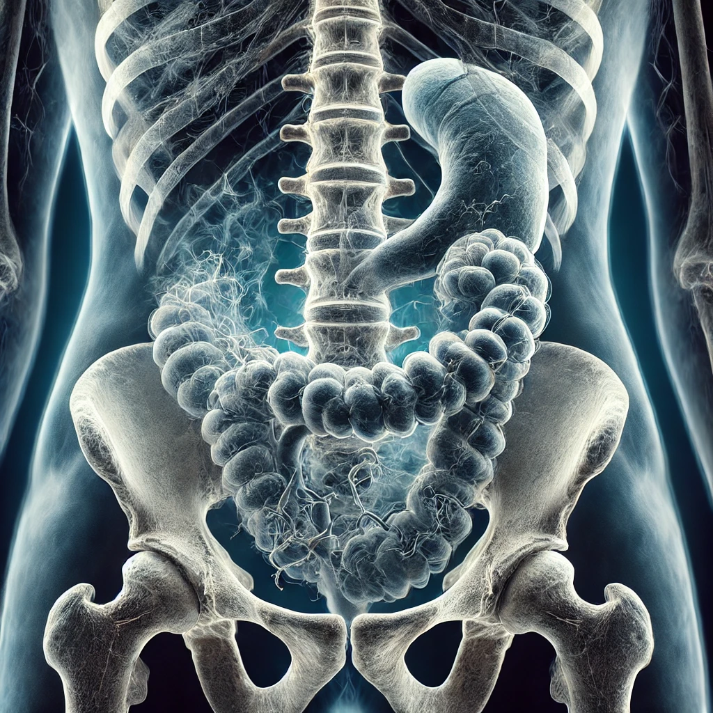
A bone density scan of the lumbar spine side also captures an image of the abdominal aorta, which supplies oxygenated blood from the heart to the abdominal organs and lower limbs.
Calcium buildup in the artery, known as abdominal aortic calcification or AAC, serves as a vital marker for predicting heart attack and stroke risks while also providing insights into brain health.
Prior human studies indicate that higher calcification levels are linked to poorer scores in verbal learning, memory, processing speed, language fluency, and overall cognitive performance. One study found that assessing AAC in midlife predicted white matter brain lesions burden twenty years later.
Given the lack of previous exploration into AAC and late-life dementia risk, researchers from Edith Cowan University in Australia studied AAC data in 968 women over a 15-year period.
At the study's onset, the participants, with an average age of 75, had varying AAC levels: low at 44.7%, moderate at 36.4%, and extensive at 18.9%. By the study's end, 15.7% had been diagnosed with or died from dementia.
After adjusting for cardiovascular and genetic factors, the results, published in The Lancet journal, revealed that compared to women with low AAC, those with moderate and extensive AAC faced double the dementia risk. On average, more than half (55%) had a two to fourfold increased risk.
Joshua Lewis, part of the research team, highlighted the common presence of bone density machines and the ease of obtaining lateral spine images during routine bone density tests. He emphasized the simplicity, cost-effectiveness, and minimal radiation exposure of these scans compared to X-rays or CT scans, suggesting their potential as a quick, safe screening tool for assessing dementia risks in older individuals.
His colleague and senior author Professor Simon Laws emphasized the link between heart and brain health, noting that modifying risk factors like diet and physical activity is crucial in dementia prevention. Early intervention is key to effecting change and maximizing impact.
Waiting for a negative finding on a bone scan as motivation for action is discouraged. The CDC recommends that seniors engage in at least 150 minutes of moderate-intensity aerobic exercise five days a week, along with two days of muscle-strengthening activities.
To enhance your diet, aim to reduce refined, processed, and sugary foods and switch to whole, natural foods. Nutritional supplements can also positively impact heart and brain health.
It's important to note that bone health not only influences your quality of life and independence as you age but is believed to be linked to your longevity by scientists.
A groundbreaking study has shown that bone density scans can predict dementia risk by identifying calcification in the abdominal aorta visible in these scans. Women with moderate to extensive plaque buildup in this artery were found to have double the risk of late-life dementia. This underscores the critical relationship between heart and brain health and stresses the importance of early intervention through diet, exercise, and lifestyle adjustments to mitigate these risks.
What is abdominal aortic calcification (AAC)?
AAC refers to the accumulation of calcium in the abdominal aorta, serving as an indicator of cardiovascular and cognitive health risks.
How does AAC impact dementia risk?
Extensive AAC is associated with a doubled risk of late-life dementia, likely due to reduced blood flow and increased inflammation.
Can bone density scans detect AAC?
Yes, lateral spine images from standard bone density tests can capture AAC, providing a non-invasive and cost-effective method to screen for dementia risk.
What lifestyle changes can help reduce AAC?
Regular aerobic exercise, strength training, a diet rich in whole, unprocessed foods, and avoiding smoking can help reduce AAC and its associated risks.
Are bone density scans safe?
Bone density scans are quick, low-cost, and involve minimal radiation exposure, making them a safe screening tool.
Susan Parker is a 49-year-old Senior Manager at a marketing firm. With two older children becoming more independent, she is now focusing on her own health and wellbeing. She’s passionate about natural and holistic health approaches, and values high-quality, trustworthy products. Susan enjoys yoga, gardening, reading, and cooking, and seeks to stay energetic and sharp while balancing a busy career and personal life.